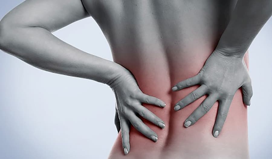
My aim in this blog is to dig a little deeper into the relatively simple structure of our skeletal muscle. Although simple, it’s invaluable to human function and it’s role is further reaching than you may think.
To start off with, skeletal muscle is only one of three types of muscle found within the body:
- Skeletal muscle forms the muscles responsible for locomotion and gross body movements.
- Cardiac muscle is only found in the heart and therefore is vital for cardiovascular function.
- Smooth muscle is found in visceral organ walls (and a few other locations) and is important for support, contractibility and elasticity.
Skeletal muscle has a greater role than we have so far given it credit for, however, it is found in the body:
- Pulling on the bones of the skeleton via tendons to create movement
- Maintaining posture
- Shielding and supporting visceral organs within the abdomen (we have heard of the importance of core strength but we often don’t think of the effect it has on abdominal viscera)
- Giving us voluntary control over swallowing, defecating an urination by opening and closing the opening of the digestive and urinary tract
- Regulating temperature within the body by releasing heat when working
- In times of dire need, when we are not consuming sufficient proteins or calories in our diet, skeletal muscle is broken down to synthesise glucose and provide energy
Your skeletal muscle is formed of bundles within bundles, the smallest unit of which is a muscle fibre. These bundles are wrapped in several layers of connective tissue, all with different names but all are continuous with one another and the collagen fibres of which combine to form the tendons or an aponeurosis (broad tendon… kind of). When muscle fibres contract they pull on these sheaths of connective tissue which transmit this pulling force on the bone.
Direct muscle attachments occur when the outermost layer of connective tissue (epimysium) connect with the periosteum or the perichondrium (outermost layer of bone or cartilage). Indirect attachments are when the connective tissue extends beyond the muscle fibres to form a tendon. Tendons are compact, which allows more space for a larger number of muscle attachments on the relatively small surface area of the bone, and are more durable, due to their high collagen content.
The perimysium, a middle layer of connective tissue, contains collagen and elastin but also blood vessels and nerves which supply the muscle fibres. This is an interesting fact to note clinically because if the muscle remains contracted (what is more commonly known as “tight”) the flow of blood and neurotransmitters within and supplying the muscle may be reduced.
Skeletal muscle fibres are a type of cell but are specialised and are different to your typical cell. They are relatively enormous. If you take a muscle in your thigh, they can span the distance between the tendons at either end of the muscle, up to 30 cm. They also have multiple nuclei (multinucleate), hundreds in fact, just internal to the plasma membrane. Sarcoplasm is the muscles cells equivalent to cytoplasm and contains large amounts of glycosomes (granules of stored glycogen) and myoglobin (red, similar to haemoglobin in that it stores oxygen). In addition to the usual organelles a muscle fibre also contains the highly myofibrils, sarcoplasmic reticulum and T tubules which we are about to delve into.
Before we go into that though, it is interesting to note that skeletal muscle fibres are incapable of dividing. New muscle fibres and created by stems cells than remain in adult skeletal muscle tissue. This is clinically important as it results in the skeletal muscle tissue being able to heal itself after an injury, which is good news as therefore conservative treatment from an chiropractor is effective.
Now to delve into the wonderful intercellular tubules that help regulate muscle contraction: the sarcoplasmic reticulum (SR) and T tubules
- Transverse Tubules (T tubules) are tubes found at each A-I band junction. The sarcolemma protrudes deep into the cell interior forming an elongated tube known as a T tubule. The run between the paired terminal cisterna of the SR to form triads. As they pass from one myofibril to the next the T tubules also encircle each sarcomere. Nerve impulses travel along the sarcolemma and as the T tubules are continuous with the sarcolemma so they conduct the impulses into the deepest regions of the muscle cell and every sarcomere. These impulses signal the release of calcium ions from the adjacent terminal cisterna. They form a network of tunnels through the muscle fibre.
- The SR is an elaborate, smooth endoplasmic reticulum. Its interconnecting tubules surround each myofibril the way a loosely crocheted jumper surrounds an arm. Terminal cisterns form cross channels at the A-I band junctions on either side of a T tubule. Closely associated with the SR are large numbers of mitochondria and glycogen granules as they are involved in the production of energy needed for muscle contraction. The SR regulates the intracellular levels of ionic calcium. It stores Ca+ and releases it when the muscle fibre is stimulated to contract.
Each muscle fibre contains many many myofibrils. They are so densely packed that mitochondria and other organelles are squeezed between them, they form 80% of cellular volume. Within myofibrils are sarcomeres- the contractile elements of skeletal muscle cells. Within the sarcomeres are myofilaments. Striations are a repeating series light and dark bands along the length of each myofibril the dark A band and the light I bands are aligned in striations and so are apparent in muscle fibres. The light bands are formed of actin and the thick filaments are formed of myosin.
The thin filaments containing actin extend across the I band and partway into the A band. The z discs anchor the thin filaments.
Actin has kidney shaped polypeptide subunits called globular actin. This is where they myosin heads bind to during contraction. Two intertwined actin filaments (looks like a double strand of pearls) forms the backbone of each thin filament.
Thin filaments also contain:
- Polypeptide strands of tropomyosin which spiral around the f-actin to stiffen and stabilise it. In a relaxed muscle fibre hey block the myosin binding sites on actin.
- Troponin a globular, three polypeptide complex. TnI is an inhibitory unit that binds to actin, TnT binds to tropomyosin and helps position it on actin, TnC binds to calcium ions.
The thick filaments contain myosin and extend the length of the A band. They are connected at the M line.
Each myosin molecule consists of two heavy and four light polypeptide chains. They have a rod-like tail and a hinge with two globular heads. The globular heads link the thick and thin filaments together during contraction to form cross bridges and swivel about their point of attachment. The tails form the middle of the thick filaments and the heads are arranged at the ends. The heads have actin and ATP binding sites and also have ATPase activity that splits ATP to generate energy.
Elastic filaments are composed of titin. It extends from the Z disc to the thick filament, forming its core, to bind to the M line. It supports the structures of the A band and helps the muscle to spring back after stretching. The part of titin that spans the I band is extensible (unfolding and recoiling). It does not impede stretching within the ordinary range of motion but resist extensive stretching.
Fun fact: When giving a protein a technical name it must include the amino acid sequence it is composed of. For your average protein this isn’t a problem, although it does give rise to some long names. In the case of titin, however, due to its relatively monstrous size (relative in the world of proteins) and to the fact that its formula is C₁₆₉ ₇₁₉H₂₇₀ ₄₆₄N₄₅ ₆₈₈O₅₂ ₂₃₇S₉₁₁, the technical name of titan is ludicrously long, forming the longest word in any language. Topping even supercalifragilisticexpialidocious.
Muscle contraction is the activation of myosin cross-bridges. Shortening only occurs if the cross-bridges generate enough tension to overcome the forces that oppose the shortening. When relaxed the thick and thin filaments overlap only at the ends of the A band. During contraction the thin filaments slide past the thick ones and overlap to a greater degree.
Each muscle is controlled by a nerve cell called a motor neuron. A single axon branches within the perimysium to form a number of fine branches, each of which ends up at an expanded synaptic terminal. Midway along a muscle fibre’s length the synaptic terminal communicates with it as part of its neuromuscular junction.
The cytoplasm of the synaptic terminal contains mitochondria and vesicles that contain acetylcholine. This is a neurotransmitter. These are chemicals released by a neuron to communicate with the surrounding cells. They change the properties of the other cells membranes, in this case permeability. The release of ACh causes changes in the sarcolemma which triggers contraction of the muscle fibre.
The motor end plate has deep folds to increase surface area and therefore maximising the number of ACH receptors.
The synaptic cleft is the narrow space in between the synaptic terminal and the sarcolemma. The motor end plate is the portion of the membrane which contains the receptors that bind to the ACh. Both the synaptic cleft and the motor end plate contain the enzyme acetylcholinesterase (AChE) which breaks down ACh. Neurons control muscle fibres by stimulating the production of an action potential (electrical impulse) in the sarcolemma.
The sequence of events goes like this:
- Arrival of an action potential at the synaptic terminal
- This triggers the release of ACh from the vesicles in thee synaptic terminal into the synaptic cleft
- ACh binds to the receptors on the motor end plate after diffusing across the synaptic cleft. This increases the permeability of the sarcolemma to sodium ions. The sudden infux of Na+ generates an action potential
- The action potential travels via the T tubules to the terminal cisternae/SR
- Return to initial state. AChE breaks down ACh.
The action potential within the T tubules triggers the release of Ca+ from the terminal cisternae. As the active sites become exposed (troponin moves tropomyosin) on the thin filaments, cross bridge interactions form and contraction begins. Due to the communicating T tubules, all terminal cisternae in the muscle fibre are affected. Therefore, contraction is a combined effort of every sarcomere on every myofibril. During contraction ACh is broken down by AChE.
In resting sarcomeres each myosin head is bound to a molecule of ADP and a phosphate group which are released from the breakdown of an ATP molecule. The energy released by this breakdown is stored in the head so, at resting, it is primed for contraction.
Contraction:
- The active site on actin is exposed when troponin binds to Ca+
- Myosin cross-bridges form and attach to the active sites on the thin filament
- The myosin head pivots towards the centre of the sarcomere, releasing the ADP and phosphate group and using the energy that was stored.
- The cross-bridges detach when they bind to another molecule of ATP
- The myosin head splits ATP into ADP and a phosphate group and stores the energy released. It is “primed” once more.
This is clinically important for us chiropractors because energy is required for RELAXATION of a muscle. We need ATP within our muscles in order to relax. Think back to the earlier comment of maintained muscle contraction, say as a result of a neurological stimulus due to pain within a joint, limiting the blood supply to that muscle (like a hose pipe being squashed within a fist). If the muscle has a reduced blood supply and the blood supply provides ATP how is that muscle going to relax? The vicious cycle formed is that due to a reduced blood supply the muscle cannot relax to improve its own blood supply … could this be how fibrosity is formed? Those chronically tight muscles that you call “tight”. Now when we apply soft tissue techniques we are essentially causing micro-damage, triggering a pain response. Handily, when the body experiences pain, it flushes that area with blood… bringing ATP back into the muscle and allowing it to relax.
Yet we find it much easier for Chiropractic to say that we are “stretching” those muscles… a feat actually not possible for our weak little thumbs.
Related Articles
- A Little Something about Skeletal Muscles
- Sports Massage Tips for Tight Muscles
- Soft-Tissue Osteopathy Techniques
- Why Athletes Use Chiropractors
- FAQs for Those New to Visiting the Chiropractor